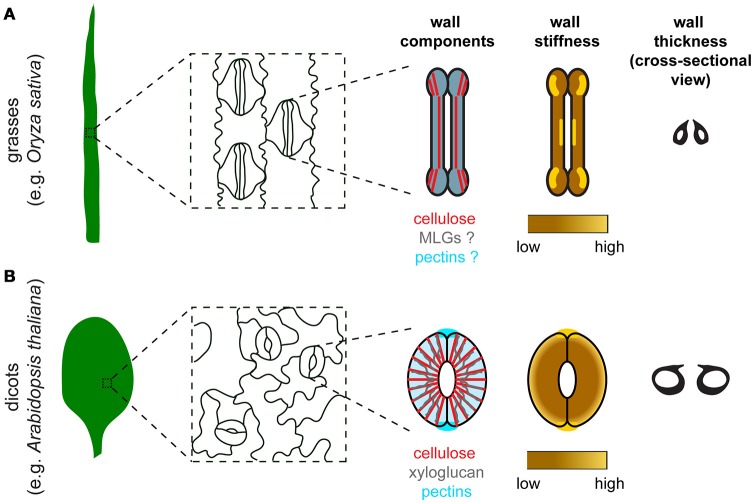
Figure 1.
Comparison of stomatal morphology, components, organization, stiffness distribution, and thickness of the guard cell wall between grasses and dicots. (A) In grasses such as Oryza sativa, guard cells are dumbbell-shaped and stomata are oriented in the same direction in the leaf epidermis. Major components of the guard cell wall in grasses include cellulose (red), mixed-linkage glucans (MLGs, gray), and pectins (blue). Cellulose organization is depicted based on polarized light microscopy data in Shtein et al. (2017). Since the organization of MLGs and pectins in guard cells is unknown, these two components are depicted to have a uniformal distribution with a merged color. Although experimental data to measure guard cell wall stiffness is missing, a speculated stiffness map is provided based on the distribution of crystalline cellulose in guard cells (Shtein et al., 2017). (B) In dicots such as Arabidopsis thaliana, guard cells are kidney-shaped and stomata are oriented randomly in the leaf epidermis. Major components of the guard cell wall in dicots include cellulose (red), xyloglucan (gray), and pectins (blue). Note that pectic HG is also enriched at the polar ends of guard cell pairs, which coincides with the high stiffness detected in the same regions. High stiffness is concentrated at the polar ends of guard cell pairs based on published AFM data (Carter et al., 2017). Differential wall thickness is illustrated in cross-sectional views of stomata in both grasses (A) and dicots (B).

