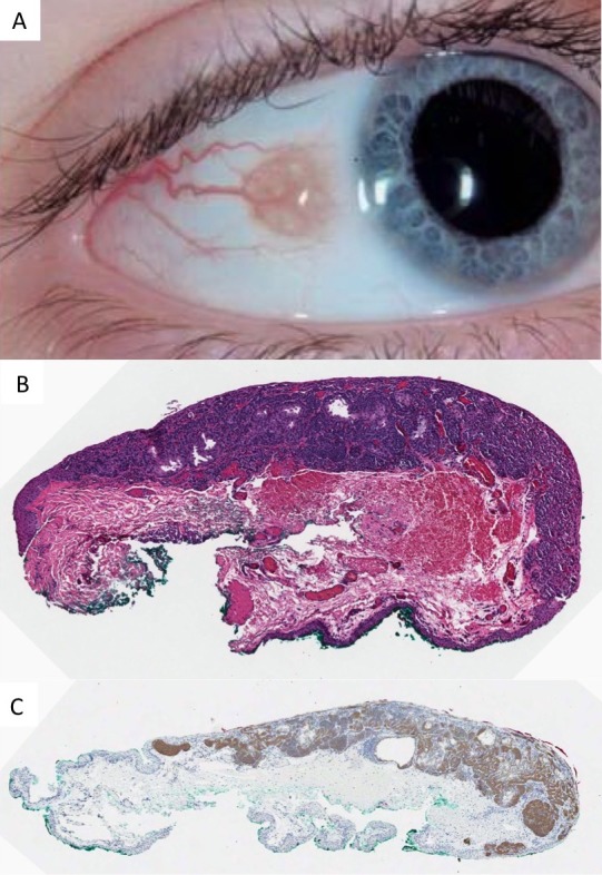
Figure 1.

Conjunctival nevus occurring in childhood. (A) Clinical photo demonstrating intrinsic cysts, diameter of 3.5 mm and amelanotic appearance. (B) Histopathology of a compound melanocytic nevus with epithelial cysts and evidence of maturation (×10, hematoxylin and eosin). (C) The junctional and subepithelial melanocytes are immunohistochemically positive for NRASQ61R (×10, peroxidase antiperoxidase).
