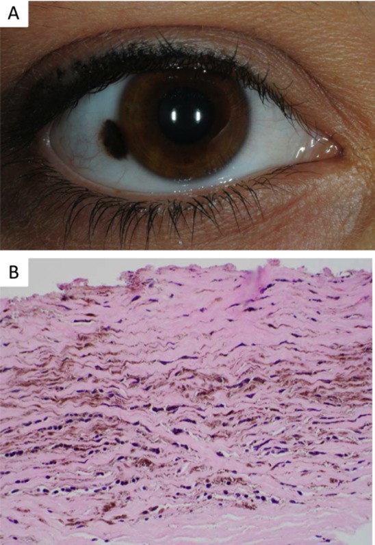
Figure 3.

Conjunctival blue nevus. (A) Clinical photo demonstrating heterochromic bulbar lesion with areas of deep melanosis. (B) Histopathology of a blue nevus characterized by pigmented slender fusiform and dendritic melanocytes in the subepithelial stroma (×10, hematoxylin and eosin).
