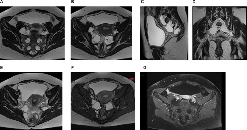
Figure 1.
MRI of giant multiple tarlov cysts. Axial (A and B), sagittal (C), and coronal (D) images: Multiformat Reformation (MPR) from 3 Dimensional T1 MRI sequence. MRI revealed 8 tarlov cysts (size: 30–63 mm) multilocular with bone erosion of the left posterior S2 vertebral body. The development of the cysts is well visible on sagittal (C) and oblique (D) views. In (A) and (B), the cysts enter the presacral space through the enlarged right and left S1–S2, S2–S3, and S3-S4 foramina. The cysts are seen as homogenous hypointense signal masses on T1-weighted image and as hyperintense signal masses on T2-weighted image. All the cysts are multiseptated: the septa (A, B: short arrows) as the nerve roots (A, long arrow) coursing within are well identified on T2 images. A cyst (E, short arrow) is seen lateral to the left ovary (E, long arrow) and another one is responsible for a compression of the sigmoid (F, arrow). (G) The multiple cysts do not enhance after gadolinium chelate administration. The cysts enter the presacral space through the right and the left enlarged S1–S2, S2–S3, and S3–S4 foramina.
Abbreviations: MPR, multiplanar reformation; 3D, three-dimensional.

