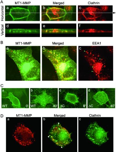
Figure 4.
Colocalization of MT1-MMP, clathrin, and EEA1 and cytoplasmic domain-dependent internalization. (A) MT1-MMP and clathrin colocalization. MDCK cells transfected with MT1-MMP (1 μg) were incubated with rabbit anti-MT1-MMP Ab at 4°C, followed by fixation and staining with mouse anti-clathrin Ab as described in Materials and Methods). MT1-MMP (green, a and d) and clathrin (red, c and f) were detected by confocal microscopy with horizontal (Upper, a-c) and vertical (Lower, d-f) scans. Two pictures were then merged and presented as b and e. Note that the expression of clathrin and MT1-MMP are heterogeneous in nature and a representative field is presented to depict clathrin inside the plasma membrane and MT1-MMP on the other side of the plasma membrane (b and e). (B) MT1-MMP in endosomes. Cells transfected as in A were fixed, permabilized, and stained with anti-MT1-MMP Ab (see Aa) or anti-EEA1 Ab (c). The same secondary Abs were used for the detections as in A. Note that the signal for intracellular MT1-MMP overwhelms its presence on plasma membrane (a). The typical ring-type endosomes are clearly visible as orange color and selectively marked by arrows (a–c). (C) Differential internalization for MT1-MMP and MT1-MMPΔC. HT1080 cells were transfected with MT1-MMP (a and b) or MT1-MMPΔC (c and d). These cells were labeled with anti-MT1-MMP Ab at 4°C for 2 h, 24 h later. The Ab was removed and cells were washed three times with cold PBS before being shifted to 37°C for commencement of the internalization program. Cells were fixed at 0 (a and c) or 40 min (b and d) and stained with FITC-conjugated goat anti-rabbit secondary Ab. Note that the wt MT1-MMP internalized into endosome-like structures in 40 min (b), whereas MT1-MMPΔC failed to be internalized appreciably (d). (D) Clathrin colocalizes with internalized MT1-MMP. HT1080 cells transfected with MT1-MMP were labeled with anti-MT1-MMP Ab and allowed to internalize for 20 min as described in C. Cells were then fixed, permeabilized, and stained with anti-clathrin Ab, followed by secondary Abs conjugated with FITC or rhodamine. MT1-MMP (red, a) and clathrin (green, c) were detected by confocal microscopy. The merged panel (b) depicts colocalization between clathrin and MT1-MMP (arrowheads).

