Figure 7.
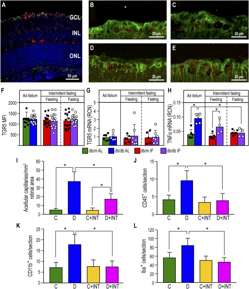
TGR5 activation using the BA agonist INT-767 prevented DR. A: TGR5 immunofluorescence staining is located in the retinal ganglion cell layer of a db/m control mouse. NeuN is used as a marker of ganglion cells. TGR5: green, NeuN: red, and DAPI: blue. INL, inner nuclear layer; GCL, ganglion cell layer; ONL, outer nuclear layer. B–E: TGR5 and NeuN staining in db/m-AL (B), db/m-IF (C), db/db-AL (D), and db/db-IF (E). F: Quantification of the fluorescence intensity of TGR5 in the retinal ganglion cell layer. Data represent means ± SD (n = 10–12). G: TGR5 mRNA expression in retinas from all cohorts. Data represent means ± SD (n = 5–7). H: TNF-α mRNA expression in retinas from all cohorts. Data represent means ± SD (n = 4–5). *P < 0.05, two-way ANOVA. I–L: Effects of INT-767 on retinas of 20-week STZ-diabetic DBA/2J mice. I: INT-767 protects from development of DR as measured by enumeration of acellular capillaries. J: INT-767 returns CD45+ cell numbers to levels seen in no diabetic mice. K: Inflammatory monocytes are reduced in the INT-767–treated mice, as shown by quantification of CD11b+ cells. L: INT-767 reduced numbers of IBA-1+ cells within the retina. C, DBA/2J mice fed with vehicle; C+INT, DBA/2J mice fed with INT-767; D, STZ/WD mice; D+INT, STZ/WD with INT-767. Data represent means ± SD (n = 6 per group). *P < 0.05, one-way ANOVA.

