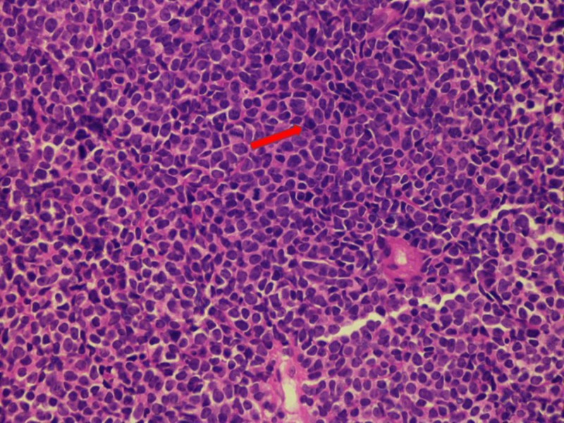
Figure 1. Histological segment of a lymph node illustrated via a hematoxylin and eosin (H&E) stain.
The H&E stain depicting nodal invasion by immature cells that are characterized by scarce cytoplasmic distribution, well-defined nuclei, and prominent chromatin. Such findings allude to the presence of a lymphoblastic lymphoma.

