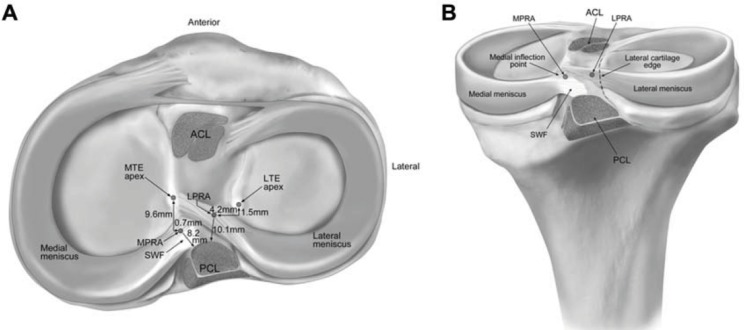
Figure 1.
Medial and lateral meniscal posterior root attachments and relevant arthroscopic bony landmarks. (A) Superior view and (B) posterior view. ACL, anterior cruciate ligament; LPRA, lateral meniscus posterior root attachment; LTE, lateral tibial eminence; MPRA, medial meniscus posterior root attachment; MTE, medial tibial eminence; PCL, posterior cruciate ligament bundle attachments; SWF, shiny white fibers of posterior horn of medial meniscus. (Reproduced with permission from: Johannsen M, Civitarese DM, Padalecki JR, Goldsmith MT, Wijdicks CA, LaPrade RF. Qualitative and quantitative anatomic analysis of the posterior root attachments of the medial and lateral menisci. Am J Sport Med. 2012;40(10):2342–7).

