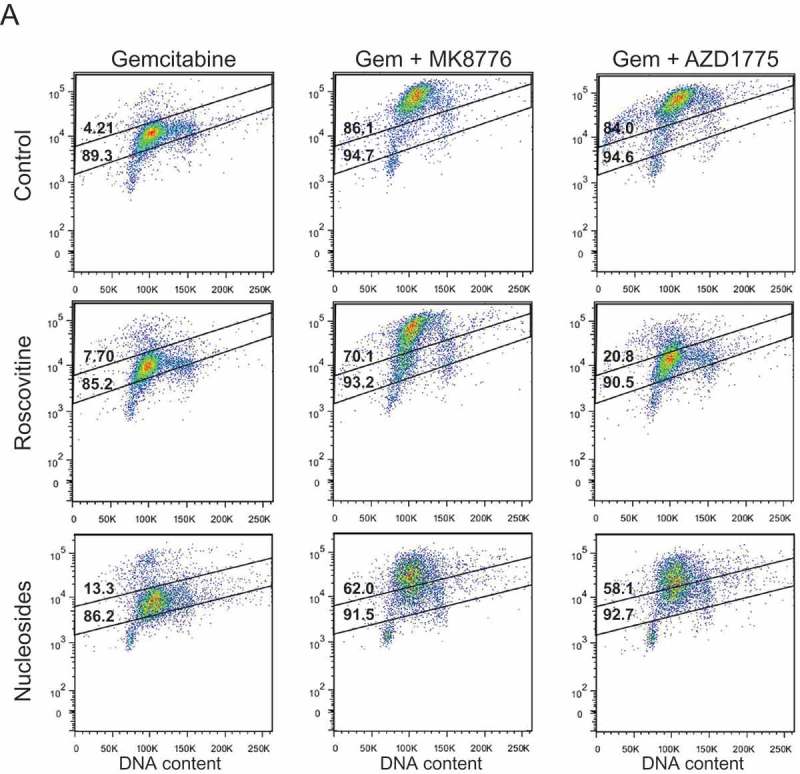
Figure 2.

The effects of roscovitine and exogenous nucleosides on MK8776 or AZD1775-mediated high-intensity γH2AX-staining in gemcitabine-treated pancreatic cancer cells. MiaPaCa2 cells were treated as illustrated in Figure 1(a) with the addition of either 20 μM roscovitine or exogenous nucleosides during inhibitor treatment, collected 30 h post-gemcitabine and assayed for γH2AX by flow cytometry (A). In each representative dot plot, the lower number is the total percentage of cells in the population considered γH2AX-positive, as defined by the larger gate, while the upper number is the percentage of cells with a high-intensity γH2AX-staining pattern, as defined by the upper gate. Cells treated with gemcitabine ± MK8776 or AZD1775 ± roscovitine or nucleosides were collected 30 or 48 h post-gemcitabine and assayed for γH2AX by flow cytometry (B – F). Data presented are the mean ± SEM from 3–8 independent experiments. Conditions that significantly attenuated high-intensity (symbols in bars) or total (symbols above bars) γH2AX staining compared to gemcitabine + either MK8776 or AZD1775 alone are indicated (*p < 0.05, one-way ANOVA). Significant differences between the effects of roscovitine, compared to the effects of nucleosides, on γH2AX staining are also indicated (δp<0.05, one-way ANOVA).
