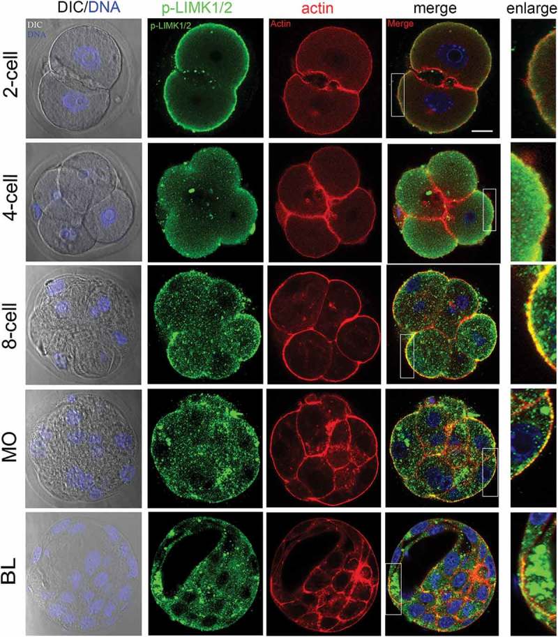
Figure 1.

The localization of p-LIMK1/2 during mouse embryo development. From 2-cell to 8-cell stages, p-LIMK1/2 was primarily localized at cortical region of blastomeres, where it co-localized with actin. And p-LIMK1/2 localized at the apical morula and blastocyst embryos. Green, p-LIMK1/2; red: actin; blue, DNA. Bar = 20μm.
