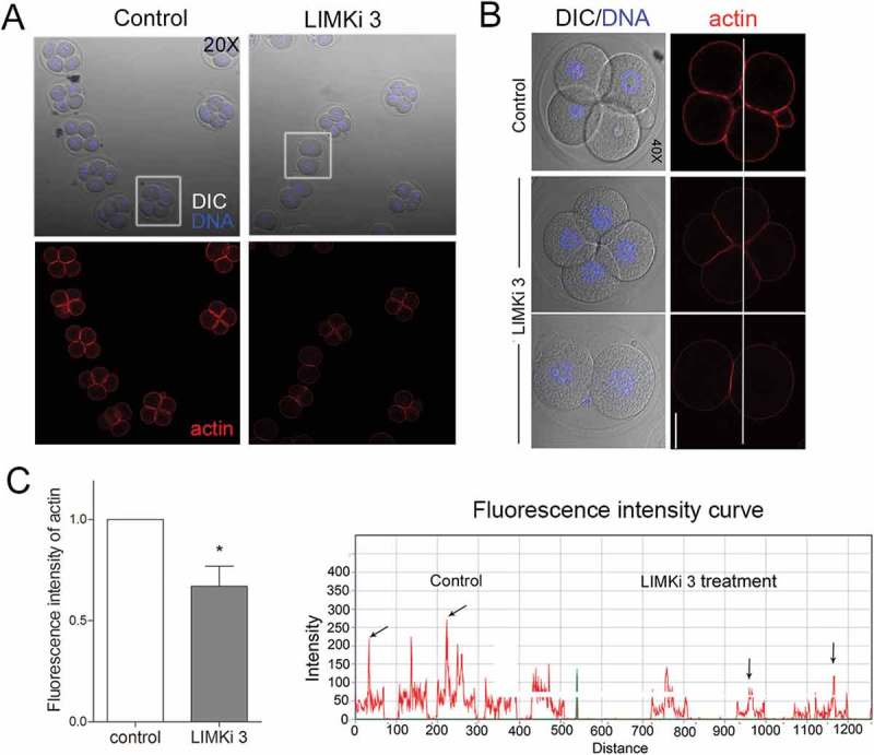
Figure 4.

Inhibition of LIMK1/2 disrupted actin assembly during early embryo development. (a) After culture for 48h, the control embryo developed to 4-cell stage, while in a proportion of LIMKi 3 treated embryos were arrested at 2-cell stage, and cortex actin signal was significantly reduced. In addition, although some embryos developed to 4-cell, the actin signal decreased compared with the control group. (b) The fluorescence intensity curve analysis showed a comparison of the actin fluorescence intensity of representative embryos (white line). (c) The fluorescent signal of actin was significantly decreased after LIMKi 3 treatment. Red: actin; blue, DNA. Bar = 20μm; *, significantly different (P < 0.05).
