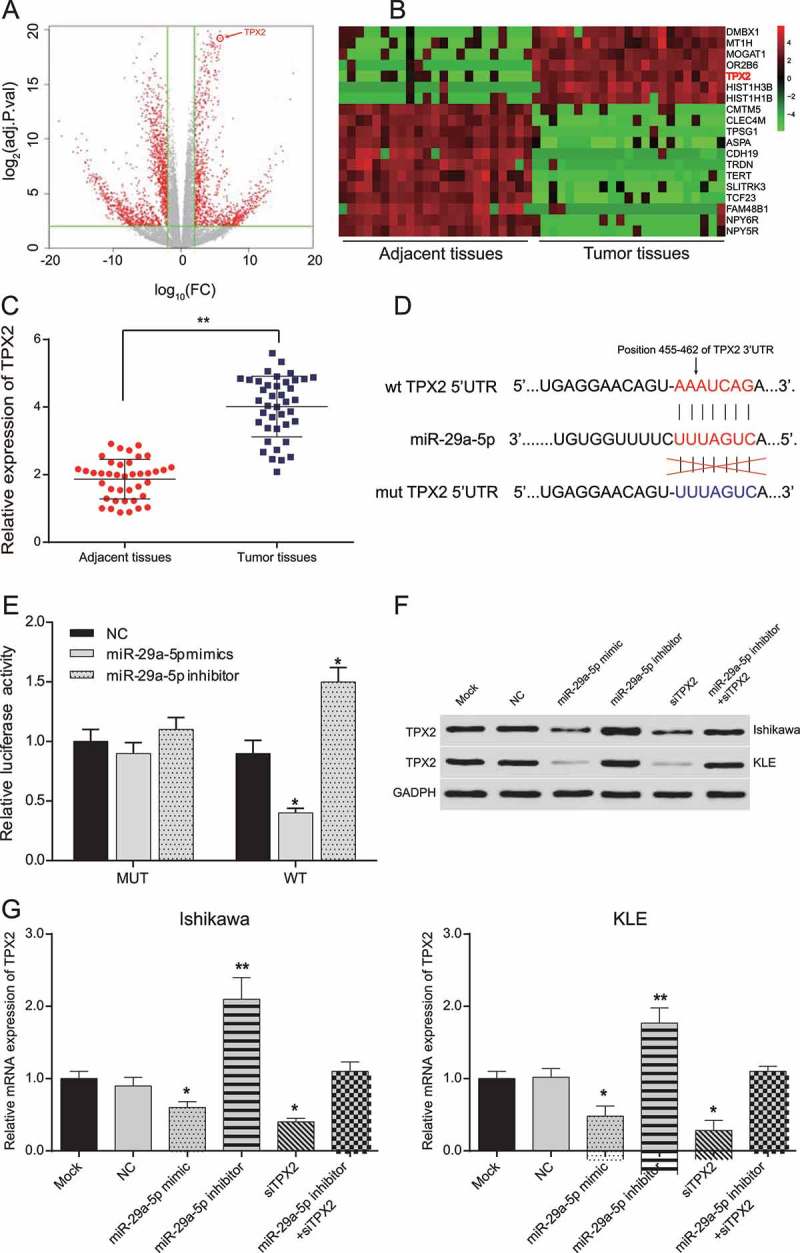
Figure 2.

MiR-29a-5p targeted TPX2 and down-regulated its expression in EC-derived cells (a-b) According to the analysis of TCGA data, differentially expressed mRNAs in EC tissues were assessed by plot and heat map. (c) A total of 40 pairs of EC tissues and adjacent normal tissues have been collected. The relative expression of TPX2 is significantly higher in tumor tissues than in adjacent normal tissues. **p < 0.01, compared to adjacent normal tissues. (d) miR-29a-5p binding sites on 3ʹUTR of TPX2 predicted by TargetScan. (e) Relative luciferase activity of EC-derived cells co-transfected with TPX2 wild-type 3ʹUTR and miR-29a-5p mimic decreased, whereas that of EC cells co-transfected with TPX2 wild-type 3ʹUTR and miR-29a-5p inhibitor increased, compared to NC group. (f) TPX2 protein expression in EC cells decreased in miR-29a-5p mimic and TPX2 siRNA transfected cells, while increased in miR-29a-5p inhibitor transfected cells, as detected by Western blotting. (g) Relative TPX2 mRNA expression in Ishikawa and KLE cells. TPX2 expression decreased in miR-29a-5p mimic and TPX2 siRNA groups, while increased in miR-29a-5p inhibitor group. *p < 0.05, compared to NC.
