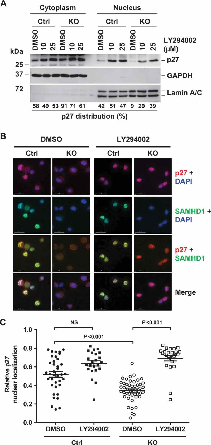
Figure 5.

PI3K inhibition reverses SAMHD1 KO-induced inhibition of p27 nuclear localization in THP-1 cells. (a) THP-1 control and SAMHD1 KO cells treated with DMSO or LY294002 (LY) for 18 h were analyzed via subcellular fractionation followed by immunoblotting for the indicated proteins. GAPDH and Lamin A/C were used as markers for cytosolic and nuclear fraction, respectively. Results are from one representative experiment of two independent assays. (b) THP-1 control and SAMHD1 KO cells were treated with DMSO or 25 µM of LY and indirect immunofluorescence was performed against p27 and SAMHD1 using specific antibodies. DAPI was used for nuclear staining. Representative images are shown. Scale bars, 15 µm. (c) Pearson’s co-localization coefficient between p27 and DAPI was quantified to determine the nuclear localization of p27 in cells. Each dot in the plot represents a cell analyzed. NS, not significant (P > 0.05). One representative experiment of three independent assays is shown.
