
Figure 6.
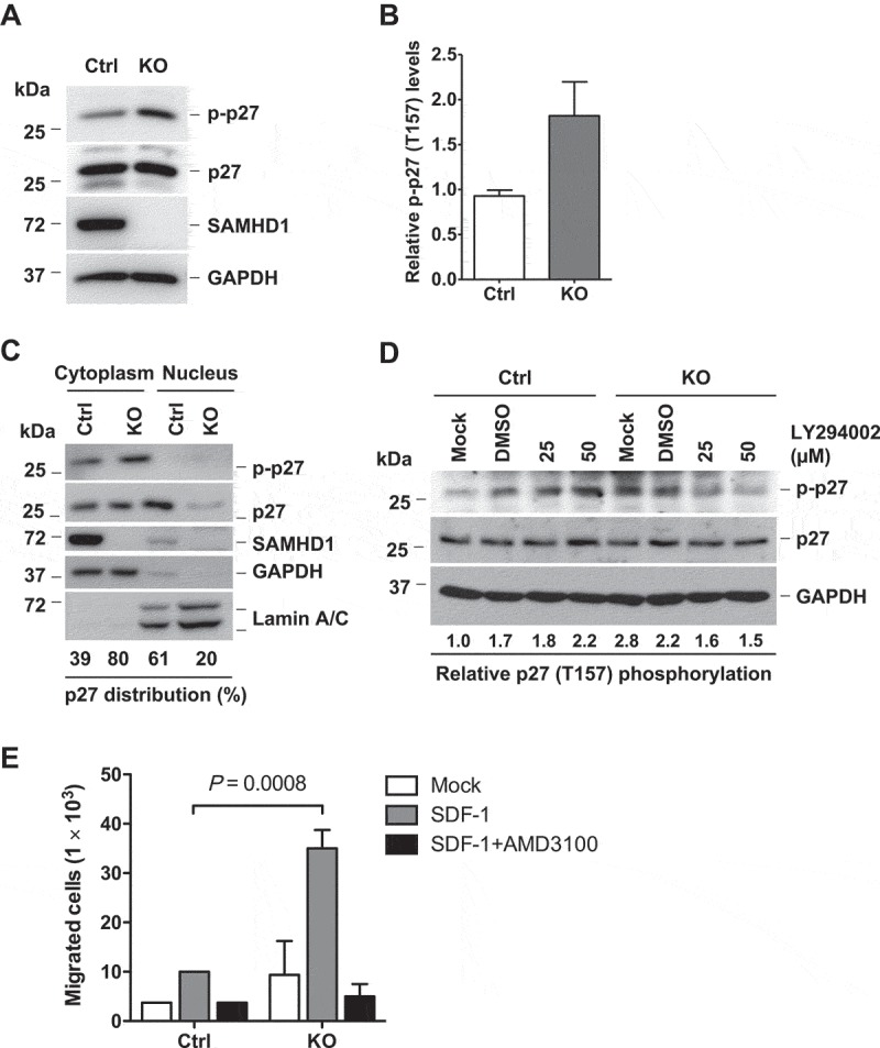
SAMHD1 KO leads to increased T157 phosphorylation of p27 via PI3K signaling and enhanced cell migration. (a) THP-1 control or SAMHD1 KO cells were analyzed by immunoblotting for the indicated protein. GAPDH was used as a loading control. (b) Densitometric analysis was performed to determine the relative levels of T157 phosphorylation of p27 (p-p27) based on the immunoblotting results of (a). (c) Subcellular fractionation in THP-1 control and SAMHD1 KO cells was performed as described in Figure 4a. (d) THP-1 control and SAMHD1 KO cells were left untreated (Mock) or treated with DMSO or LY294002 for 18 h, and p27 (total and phosphorylated form) levels were determined by immunoblotting. Densitometric analysis was performed to quantify the relative levels of T157 phosphorylation of p27 (p-p27). GAPDH was used as a loading control. (e) THP-1 control and SAMHD1 KO cells were grown in 0.1% FBS containing media with or without AMD3100 (10 µg/ml, DMSO as the vehicle control) for 90 min. Cells were then seeded in the upper chamber of a transwell plate containing media only (Mock) or media with SDF-1 in the lower chamber, and further incubated for 3 h to allow cell migration. The graph shows data from one representative experiment performed in biological duplicates. The SDF-1-induced cell migration assay represents one of three independent experiments.