Figure 2.

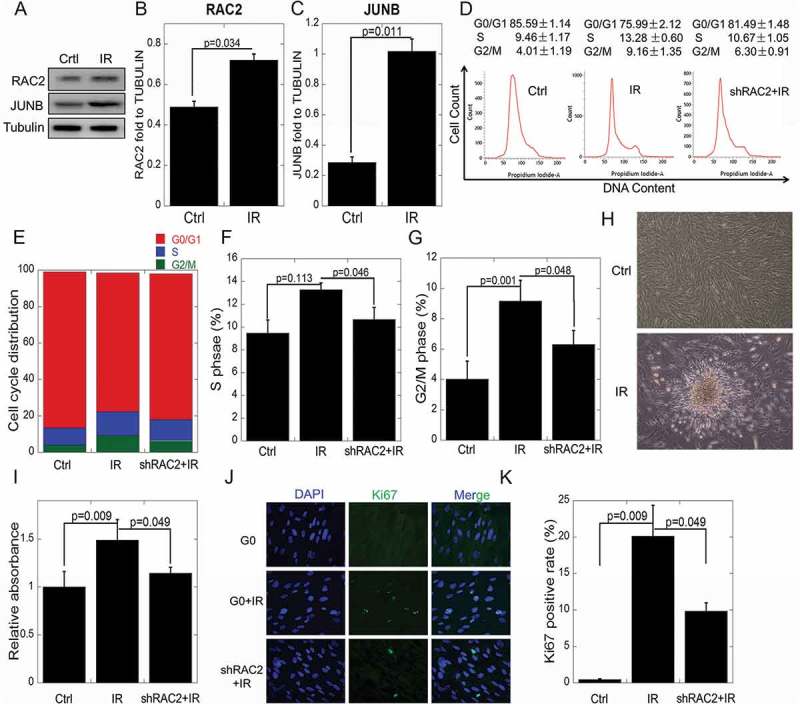
The effects of RAC2 on in vitro quiescent cell proliferation after ionizing radiation.
(a) The expression of RAC2 and JUNB were measured by western blotting. (b, c) Grayscale analyses of RAC2 and JUNB in quiescent cells and quiescent shRAC2 cells after treatment with 2 Gy of X-ray irradiation. (d–g) Flow cytometry showing significant increases in the percentages of cells in the S or G2/M phases when the cells were treated with 2 Gy X-ray irradiation. (h) The abnormal proliferation of quiescent cells after X-ray irradiation. (i) The CCK-8 assay was used to determine the viability of quiescent cells and quiescent shRAC2 cells after exposure to X-ray irradiation. (j-k) An immunofluorescent assay was used to determine the percentages of Ki67 positive quiescent cells and quiescent shRAC2 cells after exposure to X-ray irradiation. The error bars denote the mean±SE derived from three independent experiments.
