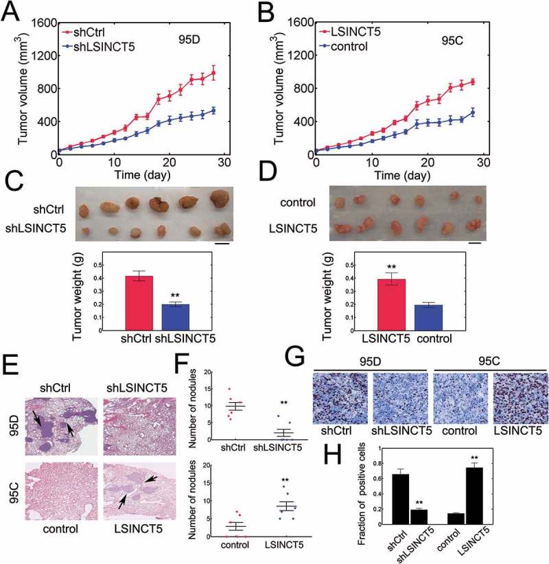
Figure 4.

LSINCT5 promotes NSCLC progression in vivo. (A) Tumor volume was quantified every 2 days for 28 days in 95D cell xenograft mouse model. 95D cells were either transfected with pLKO1-shRNA-control (shCtrl) or pLKO1-sh-LSINCT5 (shLSINCT5). **: P < 0.01. (B) Tumor volume was quantified in 95C cell xenograft mouse model. 95C cells were either transfected with pWPXL-Vec (control) or pWPXL-LSINCT5 (LSINCT5). **: P < 0.01. (C) By the end of implantation, solid tumors were resected and weighted in either 95D shCtrl or shLSINCT5 xenograft tumors. Representative images were shown (top). Tumor weight was evaluated (bottom). **: P < 0.01. (D) Tumor weight was measured in either 95C pWPXL-Vec (control) or pWPXL-LSINCT5 (LSINCT5) xenograft tumors. Representative images were shown (top). Tumor weight was evaluated (bottom). **: P < 0.01. (E) H&E staining for lung sections. **: P < 0.01. ×100 magnification. (F) The number of metastatic nodules in lung sections was quantified. (G) Ki-67 staining for tumor slides. (H) Quantification of Ki-67 positive fractions. **: P < 0.01.
