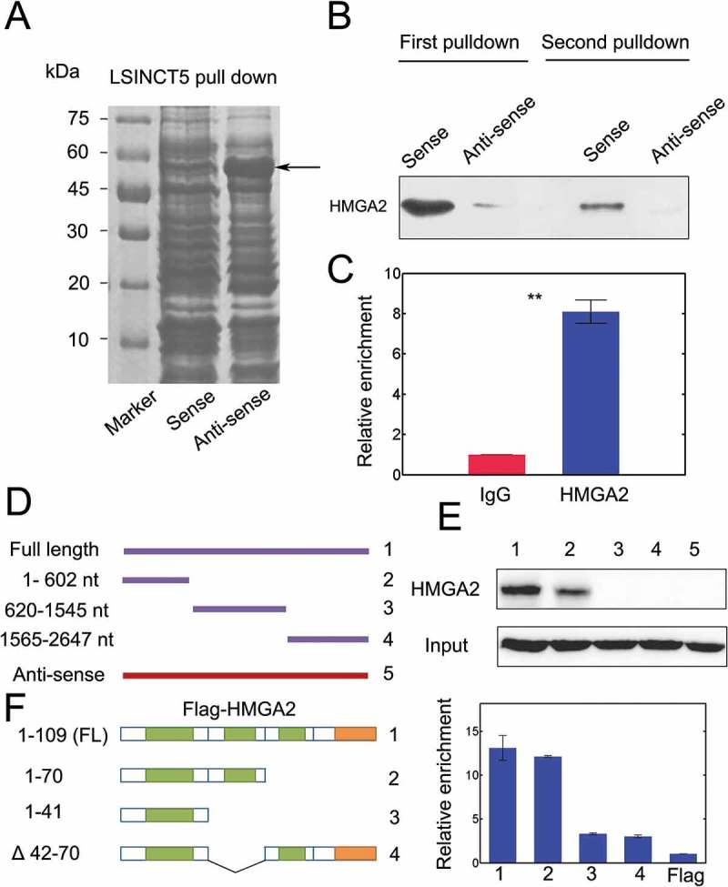
Figure 5.

LSINCT5 interacted with HMGA2 in NSCLC cells. (A) Biotin labeled LSINCT5-sense and LSINCT5 anti-sense probes were transcribed in vitro and incubated together with 95D lysates. The ~50 kD band was (arrows) excised and subject to mass spectrometry analysis. (B) Immunoblots for interaction between LSINCT5 and HMGA2 from two independent RNA pull-down assays. (C) RIP assays were done using primary antibody against HMGA2 and qPCR was used to identify LSINCT5. (D) Illustration of different truncated forms, full length (FL) and anti-sense probe of LSINCT5. (E) Immunoblots for LSINCT5 pull-down with HMGA2 using full-length (FL), various truncated forms and anti-sense probe. (F) Identification of interaction domains of HMGA2 with LSINCT5. RIP assays for LSINCT5 enrichment in 95D cells transfected with FL or different truncated forms of HMGA2. Enrichment quantification was shown on the right.
