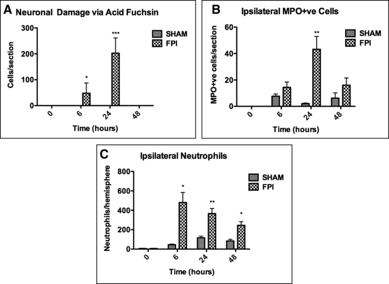
Figure 1.

Characterization of mild fluid percussion-induced brain injury. Individual hemispheres were digested 6, 24, and 48 hr after receiving fluid percussion injury (FPI)/sham procedures for flow cytometry or processed for histology. A, Neuronal damage via acid fuchsin. Significant neuronal damage was detected in the ipsilateral hemisphere 6 and 24 hr after receiving FPI. B, Myeloperoxidase (MPO) immunohistochemistry on brain sections 24 hr after sham/FPI procedure. Significant neutrophil infiltration was seen at 24 hr after FPI. C, Ipsilateral neutrophil quantification via flow cytometry. A small number of neutrophils were detected in the ipsilateral hemisphere 6, 24, and 48 hr after FPI. Data are represented as mean ± sem or sd (Fig. 1C) and were analyzed using two-way analysis of variance (n = 3–5 per group, *p < 0.05, **p < 0.01, ***p < 0.001).
