Figure 5.
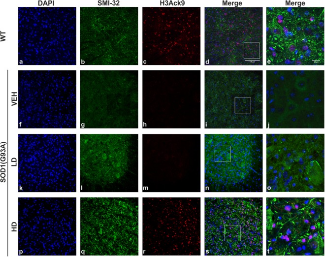
Histone 3 acetylation in the lumbar spinal cord of WT and SOD1(G93A) mice. The figure panel shows the different acetylation state of lysine 9 of histone 3 in the lumbar spinal cord of WT mice and VEH, LD and HD SOD1(G93A) groups (n = 4 per groups). The nuclei were stained in blue with DAPI (a,f,k,p) while, to identify MNs, the antibody neurofilament H was detected with SMI-32 antibody in green (b,g,l,q). The acetylation of histone 3, identified by H3Ack9 antibody in red was not present in VEH (h) and LD (m) groups compared to WT animals. The treatment with the epigenetic drugs restores the acetylation of histone 3 in HD group. Magnification 20×, scale bar 100 µm (a–d, f–i, k–n and p–s). In the last column (e,j,o,t) are shown the higher magnification of the boxed area showed in d, i, n and s respectively, scale bar 20 µm.

