Fig. 1.
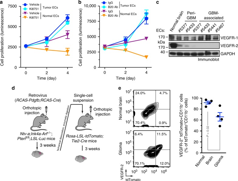
Tumor-associated ECs are resistant to anti-VEGF treatment and have diminished VEGFR-2 expression. a–c ECs were isolated from GBM tumors or peri-tumor tissues of human patients or normal brains. a, b Tumor ECs and normal brain microvascular ECs were treated with a 3 nM Ki8751 or b 10 μg/ml B20 antibody in VEGF-A-containing culture medium, and subjected to cell viability analysis (n = 3, mean ± SEM). c Cell lysates were immunoblotted. d, e The primary GBM in Ntv-a;Ink4a-Arf−/−;Pten−/−;LSL-Luc donor mice was induced by RCAS-mediated somatic gene transfer. Single-cell tumor suspension was injected into Rosa-LSL-tdTomato;Tie2-Cre mice. d Schematic approach. e Single-cell suspension isolated from normal brains or tumors were analyzed by flow cytometry. Left: representative sorting of CD11b− cells. Right: quantitative data (n = 4 mice, mean ± SEM). P value was determined by Student’s t test

