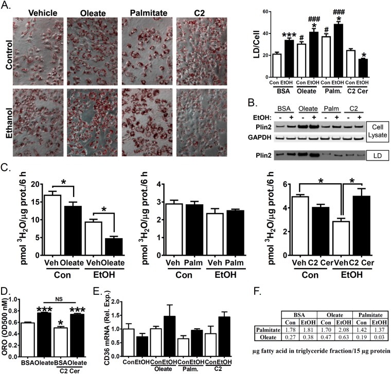
Figure 4.
Co-treatment with C2 ceramide but not oleate or palmitate reverses ethanol-mediated reduction in fatty acid oxidation. (A–F) VL-17A cells were treated for 48 h with control- or 100 mM ethanol-containing media or supplemented with BSA (vehicle control), 100 µM oleate, 40 µM palmitate or 10 µM C2 ceramide. (A) Representative images and quantitation (N = 17–20) from cells fixed and stained with Oil Red O. (B) PLIN2 and GAPDH were measured by western blotting of whole cell lysates or isolated lipid droplets. Representative cropped western blot images from different blots shown. Full-length blots are in Supplementary Figure 1. (C) Oleate oxidation was quantified by measuring 3H water liberation from 3H labelled oleate (N = 5). (D) Oil red O staining in fixed cells was quantified by elution in isopropanol followed by optical density reading at 500 nM. (E) Isolated mRNA was assayed by real time RT-PCR for CD36. (F) LC-MS/MS analysis of FA composition of triglyceride. Listed values are µg fatty acid in triglyceride fraction/15 µg protein. Data presented as mean +/−SEM. *p < 0.05 relative to BSA Con, ***p < 0.01 relative to BSA Con, ###p < 0.01, #p < 0.05 compared to BSA Con with same exogenous lipid.

