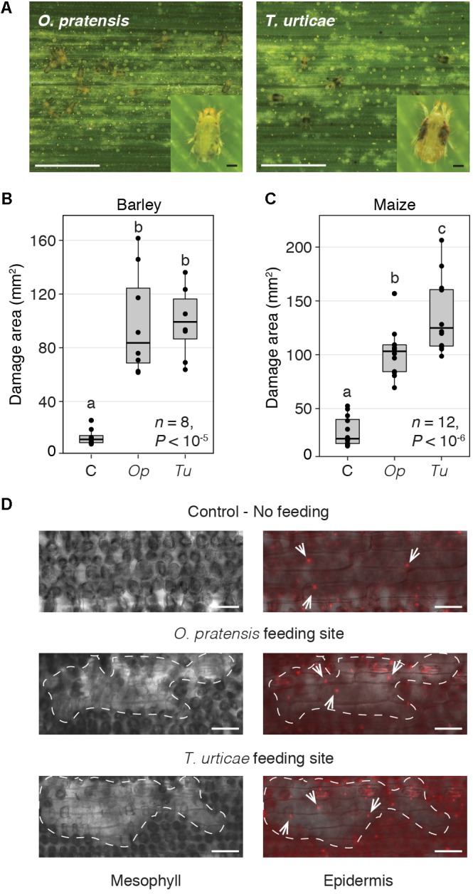
FIGURE 1.

O. pratensis and T. urticae morphology, tissue damage, and feeding strategy. (A) O. pratensis and T. urticae feeding causes light colored (chlorotic) spotting on barley leaves, as well as on maize (see also Supplementary Figure 2) (scale bars: 2.5 mm). Insets show adult O. pratensis and T. urticae females (scale bars: 100 μm). (B,C) Total area of damage on barley and maize leaf enclosures, respectively, for uninfested leaves (C, control), and those subjected to O. pratensis (Op) and T. urticae (Tu) feeding at 24 h. P-values are from an ANOVA, and letters indicate significant differences between comparisons (P < 0.05, Tukey’s HSD test; n: number of biological replicates). In the boxplots, circles represent individual data points. (D) Leaf cell damage caused by O. pratensis and T. urticae feeding on barley. Clusters of empty mesophyll cells at mite feeding sites (left) are denoted by dashed lines; a focal plane through the epidermis is shown at right with nuclei visualized by propidium iodide staining (white arrows). Scale bar: 100 μm.
