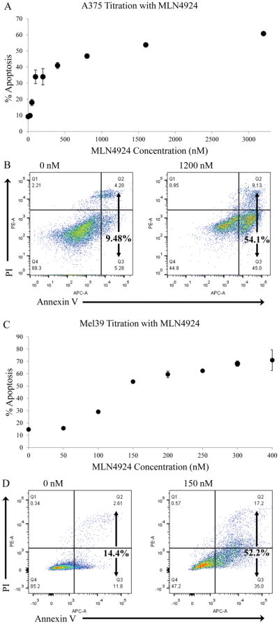
Figure 1. MLN4924 induces apoptosis in the BRAF mutant A375 and Mel39 human melanoma cell lines.

The A375 BRAF mutant (V600E) human melanoma cell line was treated for 72 hours with either vehicle or varying concentrations of MLN4924 ranging from 25 nM to 3200 nM. Following incubation, cells were harvest and Annexin V/propidium iodide flow cytometric analysis was utilized to determine the percentage of apoptotic cells (combined percentage of right quadrants). A) Graphical analysis of apoptosis (as determed by Annexin V/propridium iodide flow cytometry) of treated A375 cells. From these results, the IC50 for MLN4924 in A375 cells was determined to be 1200 nM. Data is presented as mean ± S.E.M. B) Representative flow cytometry plots of A375 cells treated with IC50 concentration of MLN4924 (1200 nM) or vehicle control (0 nM) are displayed. The same experiment for part A was repeated for the Mel39 BRAF mutant (V600E) human melanoma cell line. C) Graphical analysis of apoptosis (as determed by Annexin V/propridium iodide flow cytometry) of Mel39 cells treated for 72 hours with a titration of MLN4924 ranging from 0–400 nM. From these results, the IC50 for MLN4924 in Mel39 cells was determined to be 143 nM. D) Representative flow cytometry plots of Mel39 cells treated with IC50 concentration of MLN4924 (150 nM) or vehicle control (0 nM) are displayed.
