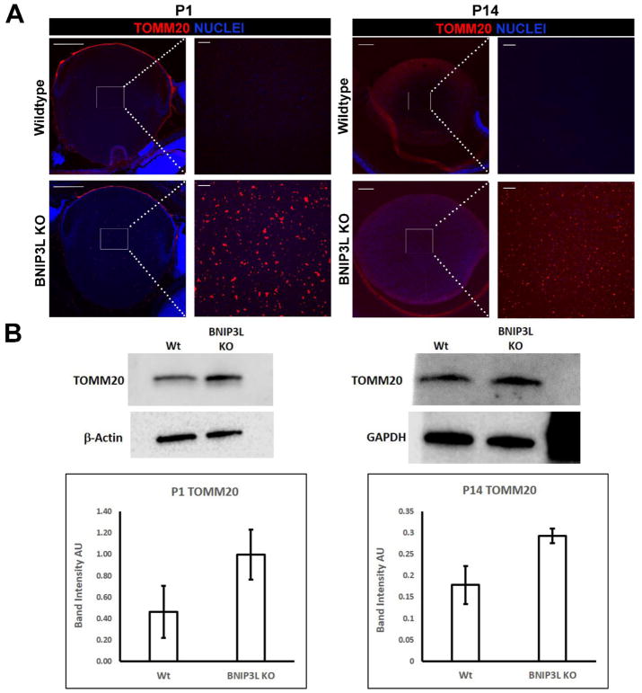
Figure 2. Mitochondria are retained in the lenses of BNIP3L knockout mice.
A. Mid-sagittal lens sections from P1 and P14, wild-type and BNIP3L KO mice immunostained, for the mitochondrial marker TOMM20 (red) and co-stained with nuclear stain DAPI (blue). Low magnification images for P1 were obtained using the 10X objective, for P14 the 5X objective was used, the scale bar for both is 200 μm. High magnification images for both P1 and P14 were taken from the center or organelle free zone of the lens and obtained using the 40X objective, scale bar 20 μm. B. Immunoblot analysis of TOMM20 protein levels in 15 μg of total protein extract isolated from P1 and P14, wild-type and BNIP3L KO lenses. Also shown are immunoblots for β-actin (P1) and GAPDH (P14) as controls for equal protein loading. Densitometric analyses of the immunoblots was plotted as TOMM20 levels relative to its loading control. Samples were run in triplicate and displayed as mean software determined arbitrary unit (AU) ± the standard deviation.

