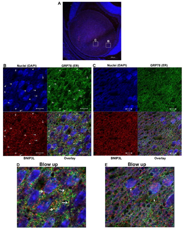
Figure 5. BNIP3L co-localizes with endoplasmic reticulum in the lens.
A. Mid-sagittal lens sections from P1 wild-type mice immunostained for the endoplasmic reticulum marker BiP/GRP78/HSPA5 (green), BNIP3L (red) and co-stained with nuclear stain DAPI (blue). A. Low magnification images for P1 were obtained using the 10X objective and the scale bar is 100 μm. B. High magnification image taken in the equatorial region of the lens. This image was obtained using the 40X objective and scale bar is 20 μm. C. Zoomed area of image B. (area indicated by box), arrows indicate co-localization of ER/GRP78 (green) and BNIP3L (red) resulting in distinct yellow puncta in the overlay image, scale bar 10 μm and D. A blow up of the overlay from C.

