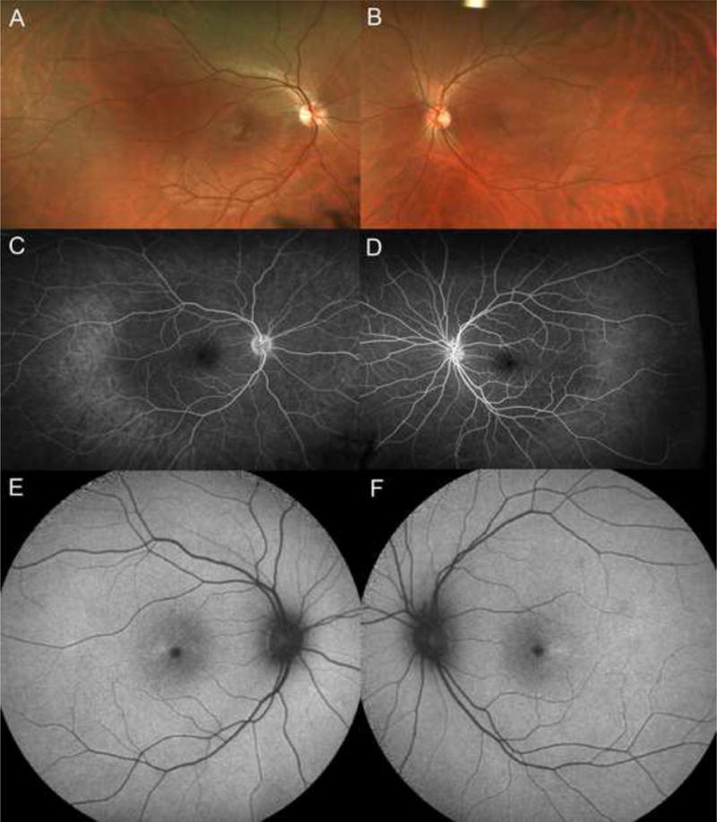
Figure 1.

A, B. Ultra-wide-field color images suggest subtle pigment mottling in the macula, with unremarkable vascular networks, optic nerves, and retinal periphery. C, D. Ultra-wide-field fluorescein angiography shows normal filling and no leakage. E, F. Fundus autofluorescence demonstrates symmetric areas of hyperautofluorescence slightly temporal to the fovea in both eyes
