
Figure 4.
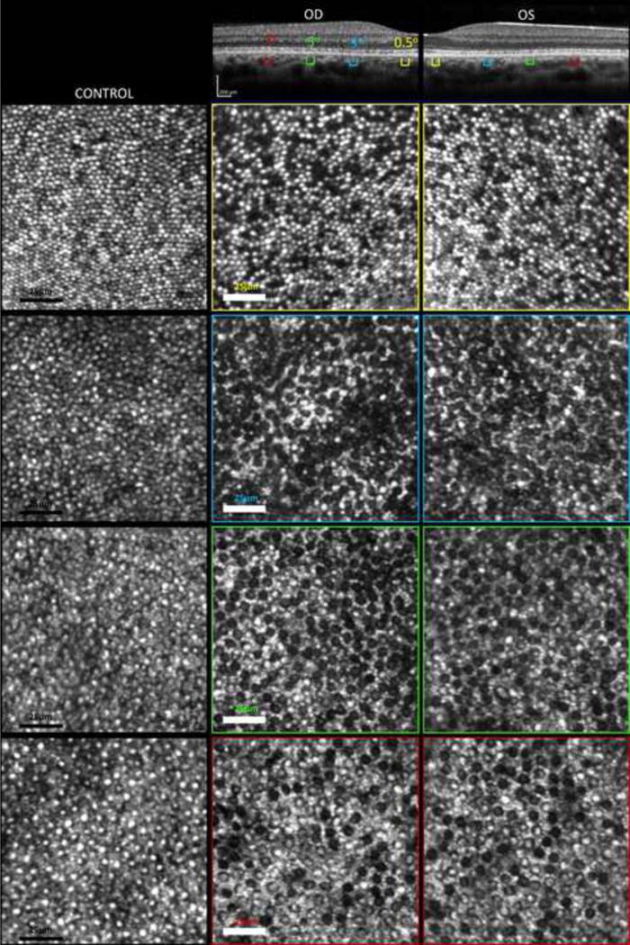
Adaptive optics scanning light ophthalmoscope images of the patient reveal multiple dark spots in the cones mosaic, not seen in the controls, consistent with loss or non-waveguiding photoreceptors at increasing eccentricities from the foveal center in both eyes (0.5, 3, 5, 7 degrees from Umbo).