
Figure 6. Examination 3 months following presentation.
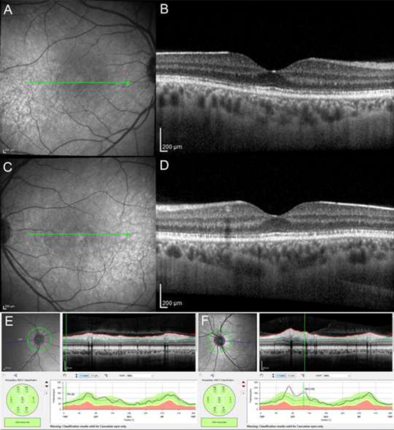
A, C. Near infrared reflectance (NIR) of the right eye (A) and the left eye (C) B, D. The OCT B-scans at the level of the corresponding area on panels A and C (green lines) evidence partial restoration of the ellipsoid zone with residual mottling. E, F. The optic nerve OCT scans demonstrate normal nerve fiber layer thickness in both eyes.