
Figure 1.
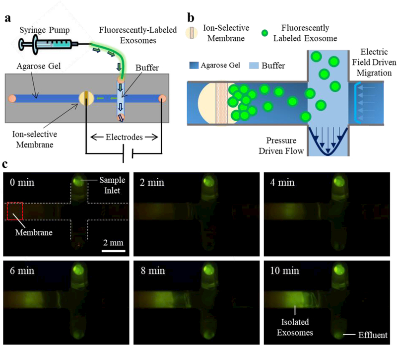
a) Schematic of the overall set-up where a syringe pump drives sample into the microchip. b) A zoomed view of the channels; as the exosomes pass through the intersection of the perpendicular channels, an electric field drives them into the gel where they concentrate at the membrane. c) A view from below the chip of the exosome isolation process with fluorescently-labeled exosomes.