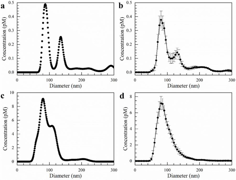
Figure 3.

Exosomes isolated directly from cell culture media using our microfluidic chip after 10 min with a flow rate of 150 µL/hr and an electric field of 100 V/cm. a) Single capture from NanoSight of cell culture media before isolation, b) Average size distribution of cell culture media before isolation. c) Single capture from NanoSight of exosomes isolated in gel. d) Average size distribution of exosomes isolated in gel. Error bars represent the standard error with n = 5.
