Figure 2.
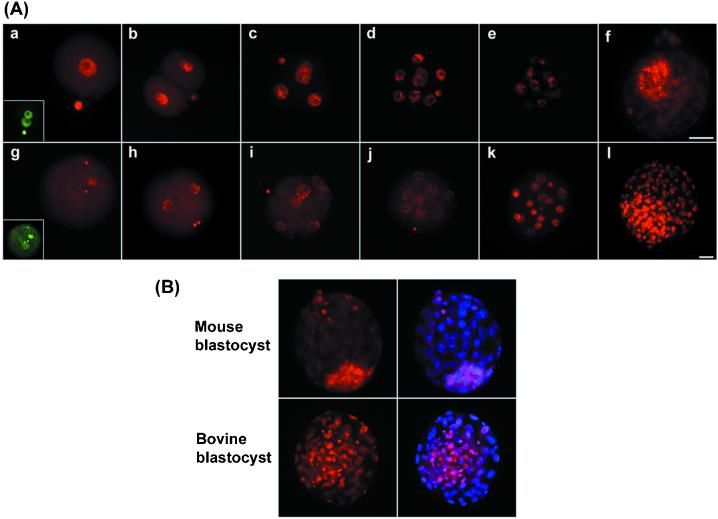
Demethylation and remethylation are conserved during preimplantation development. (A) Normal mouse (a–f) and bovine (g–l) embryos were stained for 5-methyl cytosine (red) from the zygote to the blastocyst stage. In the mouse (a–f), there is an initial loss of methylation specifically from the male pronucleus [(Inset) DNA stained to identify two pronuclei, green]. Thereafter the remaining decline in signal occurs in a stepwise fashion up to the morula stage (e). The ICM, but not the trophectoderm, has undergone de novo methylation by the blastocyst stage (f). Bovine zygotes also show loss of methylation from one pronucleus (g) followed by a further stepwise decline in methylation to the eight-cell stage (h–j). De novo methylation by the 16-cell stage results in heterogeneity with highly and moderately methylated nuclei (k) such that at the blastocyst stage (l) the ICM contains highly methylated nuclei and the trophectoderm moderately methylated ones. (B) To better define the location of the methylated nuclei images are presented with the methylation signal (red) and the merged image of the DNA (blue) superimposed on the methylation signal (pink). This superimposition of images clearly shows that in the mouse the ICM has become remethylated, but in bovine nuclei both ICM and trophectoderm are methylated.

