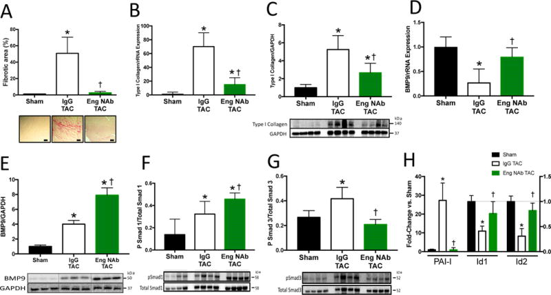
Figure 6. Neutralizing Endoglin Activity Attenuates Fibrosis and Promotes BMP9 Signaling in Heart Failure.

A) Representative histological staining for collagen and quantitation of LV fibrotic area in Endoglin neutralizing treated (Eng NAb) or IgG isotype control treated mice (IgG) after TAC. B–C) LV mRNA and protein expression of type I collagen after TAC. D) LV mRNA expression of BMP9. E) Representative immunoblots and quantitation of LV BMP9 expression normalized to GAPDH expression. F–G) Representative immunoblots and quantitation of phosphorylated Smad 1 (pSmad1), phosphorylated Smad 3 (pSmad3), total Smad 1, and total Smad 3. H) LV mRNA expression of inhibitor of differentiation 1 and 2 (Id1,Id2), and plasminogen activator inhibitor-1 (PAI-I). Scale bars 40 um. Data expressed as fold change vs. Sham. p<0.05: *, vs. Sham; †, vs. IgG TAC.
