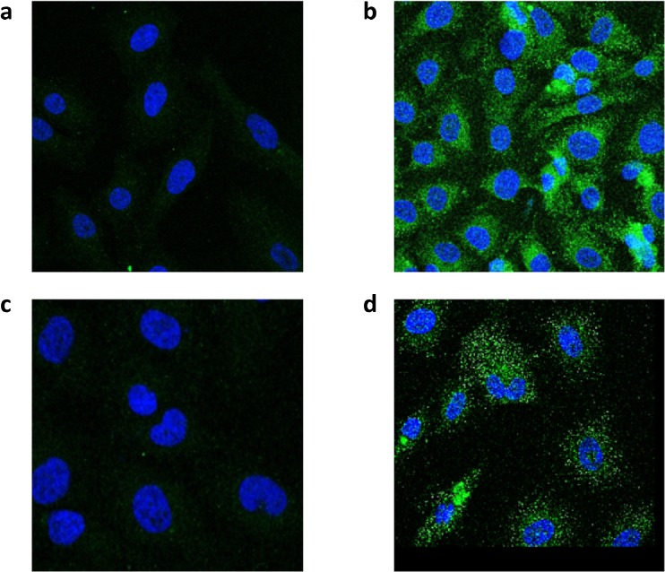
Fig. 4.
Immunostaining using keratin 9 and Hsp70 mouse monoclonal antibodies and visualized by confocal microscopy. T24 human bladder cancer cells were processed as described in “Material and methods.” The negative controls without primary antibodies (a, keratin 9) and (c, Hsp70) show slight background staining resulting from autofluorescence of the secondary antibody. The nuclei were stained with DAPI. Perinuclear distribution of keratin 9 (b) and punctate distribution of Hsp70 (d)

