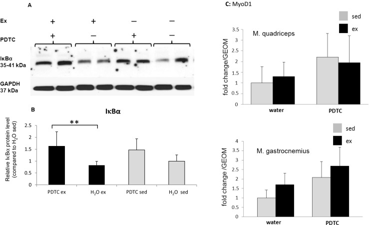
Fig. 1.

IκBα concentrations in individual skeletal muscles. a Representative Western blot analysis of IκBα concentrations in the M. tibialis anterior of sedentary and trained, PDTC-treated and control mice as indicated. GAPDH levels were analyzed as a control for equal loading and served as a standard for normalization of quantification. b Densitometric quantification of IκBα levels. Data are displayed as means ± SD (*p < 0.05, **p < 0.01; n = 8). c qPCR analysis of the gene encoding MyoD1 in Q and G. Data are displayed as geometric means (GEOM) ± SD (n = 8)
