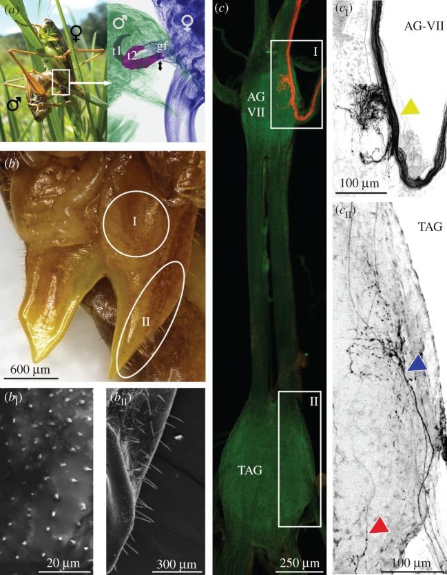
Figure 1.
Sensory sensilla at the female's genital fold. (a) A mating couple (left) and X-ray micro-CT close-up view (right) of male and female genitalia during copulation (details: [27,28]). During mating, the male's paired titillators (t1, t2; highlighted in purple) rhythmically tap (black arrows) against the female's genital fold (gf). (b) Dorsal top view of the female's genital fold. Electron microscopy scans show median area of the genital fold (bI) covered with short cuticle pegs and outer rim area (bII) with longer sensilla (details: [27]). (c) Confocal scan of TAG and last unfused abdominal ganglion (AG-VII) with fluorescence labelling (red) of sensilla from the genital fold. (cI) Close-up of afferent axon bundle entering AG-VII (yellow arrowhead). (cII) Close-up of the labelled afferent axons in the TAG (blue and red arrowheads).

