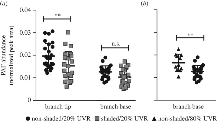
Figure 1.
Normalized abundances of platelet-activating factor (PAF; mean ± s.d.) in Acropora yongei corals depending on the location on (a) a branch (tip or base) and light conditions (shaded and non-shaded). To test the effect of tissue growth on PAF production, growing branch tips were compared to non-growing branch bases grown under non-shaded/20% UVR (n = 25 for both, black circles) and shaded/20% UVR (n = 23 for both, grey squares) conditions (to create high and low growth in the tips, respectively). To test the effect of elevated UVR exposure on PAF production, UVR was increased by 4X and the PAF content was (b) compared between non-growing bases grown under non-shaded/80% UVR (n = 11, black triangles) and the non-shaded/20% UVR conditions from (a). Data in (a) were log-transformed and compared with an analysis of variance. Data in (b) were compared with a Mann–Whitney U-test. Asterisks denote the level of significance (n.s., **p < 0.01).

