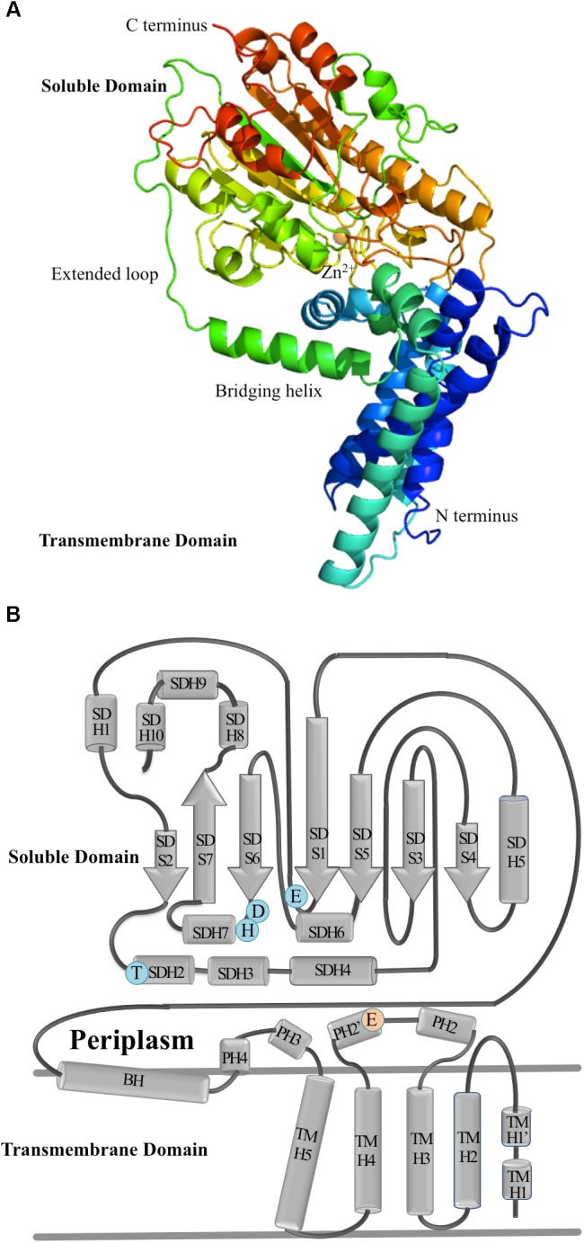
FIGURE 4.

The molecular structure of EptA. (A) A ribbon diagram showing the secondary structure elements of EptA. Coloring from blue to red corresponds to the N- and C- termini, respectively. The orange sphere denotes the position of the Zn2+ ion. (B) Topology diagram of EptA. The cylinders correspond to helices and the arrows correspond to the strands. Key amino acid residues making up the active site of the enzyme are shown in blue and orange circles. Blue circles correspond to residues which coordinate the Zn2+ ion and the orange circle corresponds to a conserved glutamate residue proposed to interact with the amine group of PEA.
