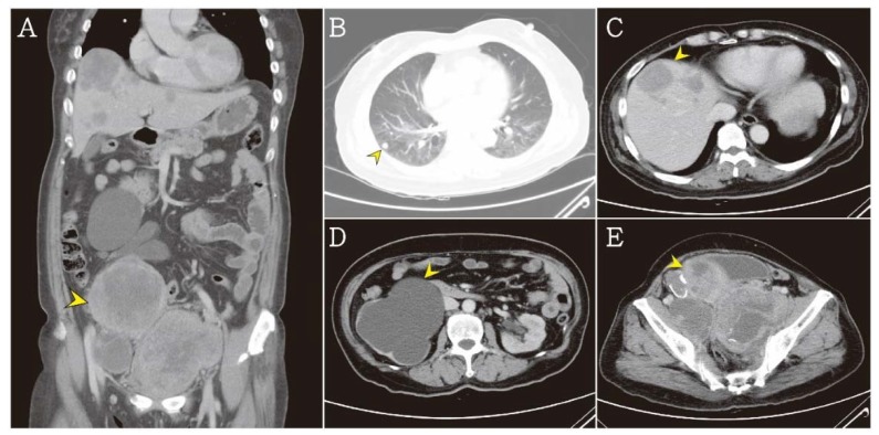
Figure 3.
(A) The abdominal computed tomography revealed a huge heterogeneous density mass (arrow head). (B) Multiple pulmonary nodules were noted in lung field (arrow head). (C) Multiple heterogeneous hepatic tumors were noted (arrow head). (D) Severe hydronephrosis at right kidney were noted due to tumor compression (arrow head). (E) Three parts of heterogeneous pelvic tumors were noted about 9.4 × 8.6 × 9.3 cm3, 7.1 × 6.1 × 6.0 cm3, 11.1 × 7.5 × 9.1 cm3 (arrow head).

