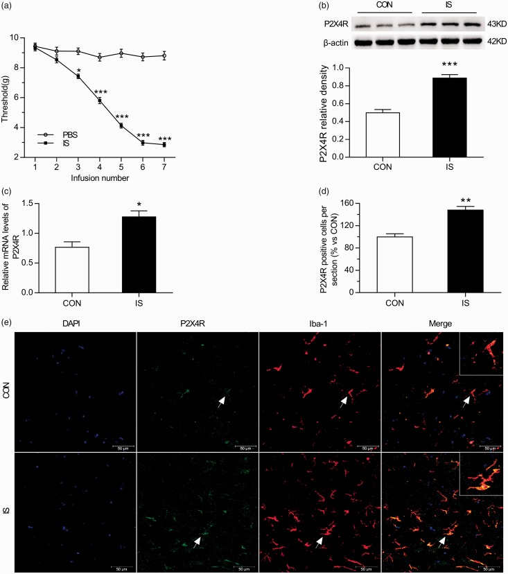
Figure 2.
Trigeminal sensitivity and P2X4R expression in the TNC increase with repeated dural stimulation. (a) Decreased baseline mechanical thresholds following repeated dural stimulation with IS. The basal mechanical thresholds of periorbital decreased in the IS group and were significantly different from the PBS group after the second infusion. (b) WB for P2X4R expression in the TNC (upper panel) revealed that P2X4R protein level was upregulated in the IS group compared with the CON group. Quantification (lower panel) of WB experiments normalized to actin control indicated an approximate twofold increase in IS group. (c) RT-PCR for P2X4R in TNC revealed that P2X4R mRNA level was significantly increased in the IS group compared with the CON group. All data were normalized to GAPDH controls. (e) Co-localization images of P2X4R (green) with Iba-1 (red) in the TNC reveal increased microglial activation and P2X4R expression in the IS group compared with the CON group. Blue indicates DAPI immunoreactivity, green indicates P2X4R immunoreactivity, red indicates Iba-1 immunoreactivity, and yellow indicates the merged signal. (d) Histogram showed the statistical result of P2X4R expression in TNC (e). Data represent the means ± SEM. Statistical analyses in (a) were performed by two-way ANOVA, followed by a Tukey test; *P < 0.05, ***P < 0.001 (n = 12 per group). Statistical analyses in ((b), (c) and (d)) were performed by independent samples T test; *P < 0.05, **P < 0.01, ***P < 0.001 (n = 6 per group in (b) and (c); n = 3 per group in (d)). Arrows indicate cells shown in the top right corner of images at approximately 4X magnification. Scale bar = 50 μm.
RT-PCR: real-time polymerase chain reaction; WB: western blot; IS: inflammatory soup; PBS: phosphate-buffered saline; CON: control.

