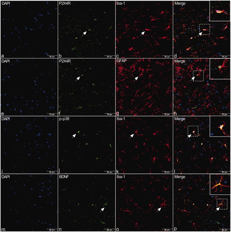
Figure 4.
Double immunostaining of P2X4R (b, f, green), p-p38 (j, green), and BDNF (n, green) with Iba-1 (c, k, o, red), a microglial marker, or GFAP (g, red), an astrocyte marker, in the TNC of rats. (a) to (h) Immunofluorescence (IF) in TNC from rats revealed that P2X4R is present in Iba-1-but not in GFAP-immunoreactive cells (shown by arrows in a–h). (i) to (p) Similar to P2X4R, IF in TNC revealed that p-p38 and BDNF staining were observed primarily in Iba-1-positive cells (shown by arrows in i–p). Arrows indicate cells shown in the top right corner of images at approximately 4X magnification. Scale bar = 50 μm.
BDNF: brain-derived neurotrophic factor; DAPI: 6′-diamidino-2-phenylindole; P2X4R: P2X4 purinoceptor; GFAP: glial fibrillary acidic protein.

