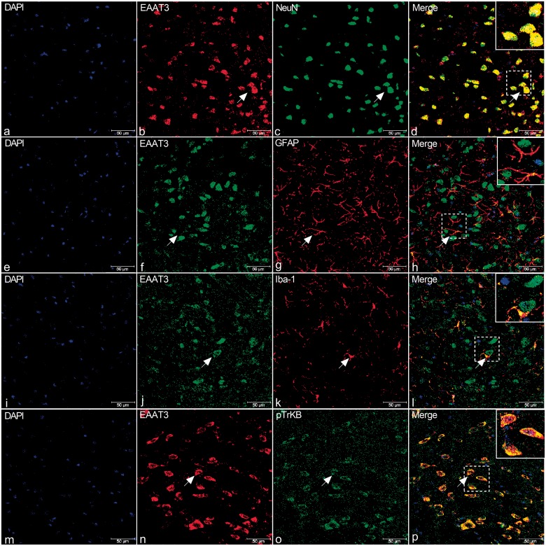
Figure 5.
Double immunostaining of EAAT3 (b, n, red; f, j, green) with NeuN (c, green), GFAP (g, red), Iba-1 (k, red), and p-TrkB (o, green) in the TNC of rats. (a) to (l) Immunofluorescence (IF) in TNC of rats revealed that EAAT3 is present primarily in NeuN-positive cells (shown by arrows in a-d). There was relatively less expression in Iba-1-positive cells (i) to (l), and expression in GFAP-positive neurons was not detected (e) to (h). (m) to (p) IF in TNC of rats revealed that EAAT3 (n) and TrkB (o) co-expressed (yellow, p). Arrows indicate cells shown in the top right corner of images (d, h, l, p) at approximately 4X magnification. Scale bar = 50 μm.
DAPI: 6′-diamidino-2-phenylindole; EAAT3: excitatory amino acid transporter 3; GFAP: glial fibrillary acidic protein; TrkB: tyrosine receptor kinase B.

