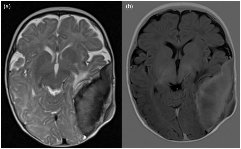
Figure 2.
On magnetic resonance imaging (MRI), the soft tissue component showed (a) mildly hypointense signal on T2-weighted images and (b) hyperintense signal on T1-weighted images. Mass effect upon the underlying left temporo-parietal lobes was observed with mild vasogenic oedema within the left temporal lobe.

