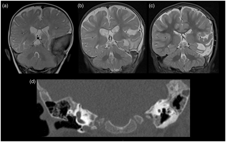
Figure 5.
Coronal T2-weighted images (a) before surgery, (b) one month after surgery and (c) one year after surgery demonstrated an area of residual sclerosis within the left petrous temporal bone which subsequently showed interval reduction in extent on follow-up at one year. Coronal computed tomography (CT) image (d) showed the area of residual sclerosis within the left petrous temporal bone one month after surgery.

