Figure 1.
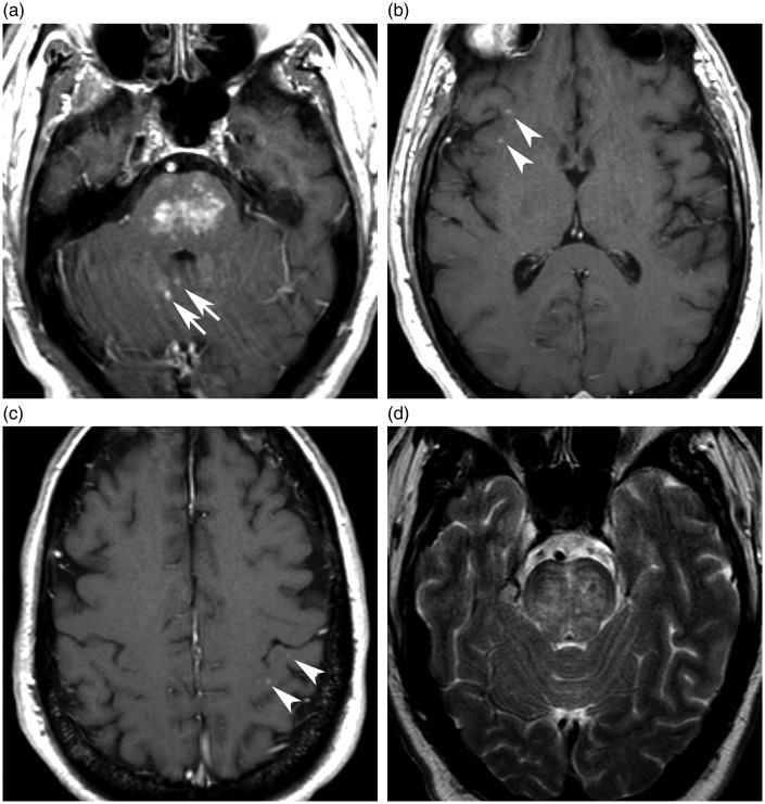
Selected images from MRI brain exam obtained at presentation, ECD of the CNS mimicking imaging findings of CLIPPERS. (a) Axial T1-weighted post-contrast image through the pons shows small, discrete enhancing lesions coalescing in the central pons. Additional cerebellar lesions (arrows). (b–c) Axial T1-weighted post-contrast images through the cerebral hemispheres show additional punctate lobar subcortical lesions (arrowheads). (d) Axial T2-weighted image through the pons shows transverse bands of edema, and mild swelling, a pattern of CLIPPERS reported by Taieb and colleagues.3
CLIPPERS: chronic lymphocytic inflammation with pontine perivascular enhancement responsive to steroids; CNS: central nervous system; ECD: Erdheim–Chester disease MRI: magnetic resonance imaging.

