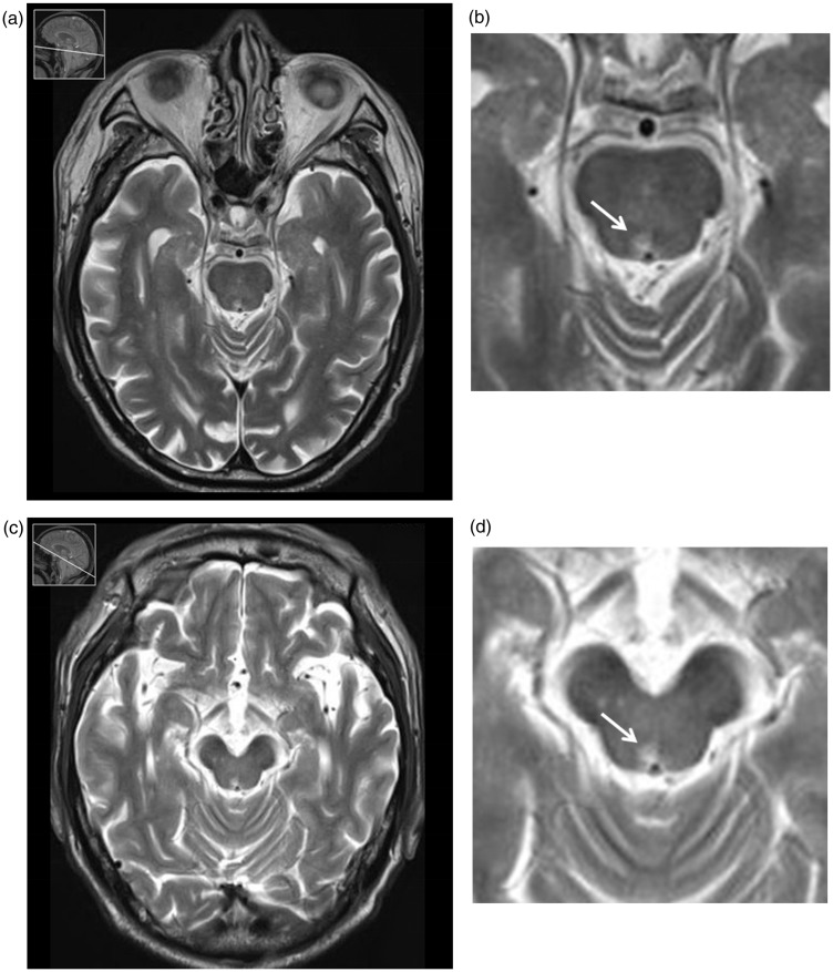
Figure 11.
A 79-year-old male presented with medial longitudinal fasciculus (MLF) syndrome due to upper pontine tegmental infarction. (a) T2-weighted brainstem vertical plane at the level of the upper pons. (b) The lesion is shown at the level of the upper pons; therefore, MLF syndrome can be diagnosed. (c) T2-weighted anterior commissure-posterior commissure plane at the level of the cerebral peduncles. (d) The lesion is shown at the level of the midbrain; therefore, the diagnosis of MLF syndrome might be difficult.

