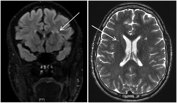
Figure 3.
Fluid-attenuated inversion recovery (left) and T2-weighted (right) magnetic resonance imaging scans for patient 013. Using the algorithm, both radiologists confirmed a non-hypomyelination pattern. Multifocal white matter signal abnormalities (WMSAs) were noted (see white arrows for a focus). These WMSAs progressed to confluence over seven years of longitudinal scans. After progressing through the algorithm, both radiologists used this information to arrive at category 7.17 (true positive outcome). This category correctly included the confirmed differential diagnosis of Fabry disease.

