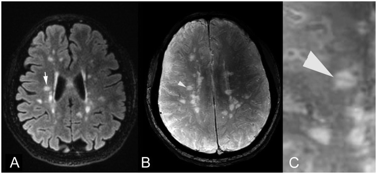
Figure 1.
Axial (a) FLAIR MR image shows a T2 hyperintense MS periventricular lesion (arrow). On corresponding (b) SWI image and (c) magnified SWI image, the central vein sign was identified as a linear signal void indicating a small central vein within the MS lesion (arrowhead). FLAIR: fluid-attenuated inversion recovery; MR: magnetic resonance; MS: multiple sclerosis; SWI: susceptibility-weighted imaging.

