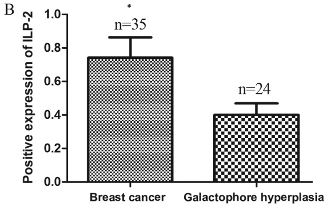
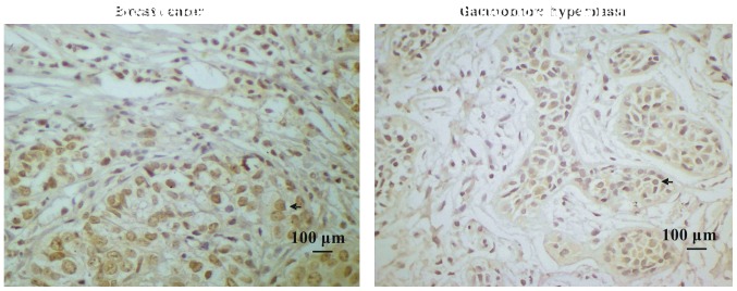
Figure 1.

(A) Immunohistochemistry of ILP-2 expression in tissues. Samples of 35 breast cancers and 24 galactophore hyperplasias were analyzed. Scale bar, 100 µm. (B) The positive rate of breast cancers and galactophore hyperplasias was 62.9 and 25%, respectively. Groups were compared by conducting χ2 test and t-test. *P<0.05 vs. the galactophore hyperplasia samples; n=59.

