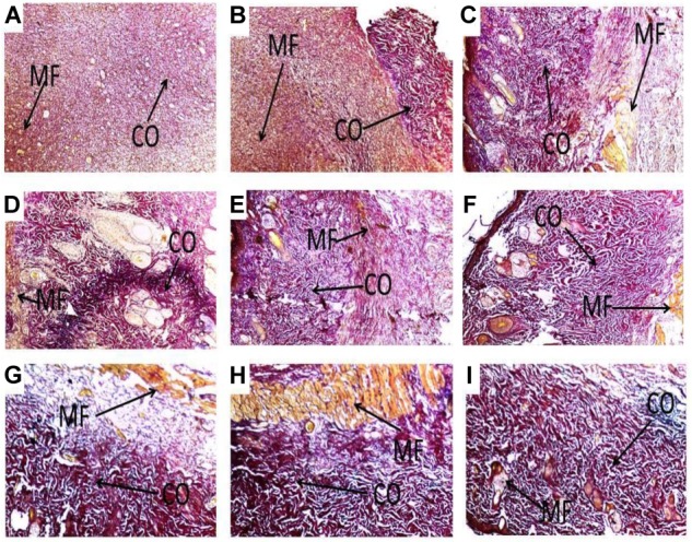
FIGURE 5.

Histological images showing influence of PLE and geraniin on collagen production in both PLE and geraniin-treated and untreated wound tissues. Full thickness wounds were created on the dorsal region and topically treated with PLE, geraniin, silver sulphadiazine, vehicle (cream only). Wound tissues were excised 13 days post wounding and sectioned. Serial sections were stained with Verhoeff–Van Gieson stain and evaluated microscopically for degree collagen deposition (Van Gieson stain ×400). Wound tissues were blindly examined by a pathologist. (A) Untreated wound tissues, (B) vehicle-treated (aqueous cream only) wound tissues, (C) 1% w/w silver sulphadiazine-treated wound tissues, (D) 0.25% w/w PLE-treated wound tissues, (E) 0.5% w/w PLE-treated wound tissues, (F) 1% w/w PLE-treated wound tissues, (G) 0.1% w/w geraniin-treated wound tissues, (H) 0.2% w/w geraniin-treated wound tissues, and (I) 0.4% w/w geraniin-treated wound tissues. CO, collagen fibers; MF, muscle fibers.
