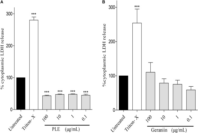
FIGURE 8.
Influence of PLE (A) and geraniin (B) on LDH release from HaCaT-keratinocytes. HaCaT-keratinocytes were seeded into 96-well plates and incubated to achieve 90% confluency. Cells were then treated with PLE (0.1 to 100 μg/mL) and geraniin (0.1 to 100 μM). Fetal calf serum (1% FCS) was used as positive control. The absorbance was measured at 490 against 690 nm. PLE, aqueous extract of the aerial parts of P. muellerianus. Values are mean ± SEM, n = 3. ∗∗∗p < 0.001 compared to untreated cells (one-way ANOVA followed by Dunnett’s post hoc test).

